Anterior Shoulder Tendon Anatomy / Self Muscle Massage pt 14- Anterior Shoulder | Muscles ... : Shoulder anatomy for ultrasound evaluation.. The conjoint tendon can be describe as a layer of connective tissue which connects the pelvis to. Latarjet procedure performed more commonly than bristow. Specifically, the four rotator cuff muscles include the following Adducts and medially rotates arm; An anterior projection of the scapula.
The name gets its origin from its structure which is often conjoined or continuous with. Majority of anterior shoulder dislocations are due to trauma. The tendon crosses anterior to the ankle joint and attaches to the. It usually results from your tendon being. Transfer of coracoid bone with attached conjoined tendon and ca ligament.
Evaluation of the Shoulder - Musculoskeletal and ... from www.merckmanuals.com The primary function of the knee is to hinge at the lower extremity. Knee function is determined in large part by the anatomy of the joint. Shoulder anatomy for ultrasound evaluation. Muscles of the anterior shoulder. Anterior aspect of the left elbow, showing superficial structures. The ri is a triangle shaped region between the supraspinatus and supscapularis tendons. The long biceps tendon arises from the supraglenoid tubercle and partly from the superior glenoid labrum (7a). Learn vocabulary, terms and more with flashcards, games and other study tools.
Related online courses on physioplus.
Biceps brachii origin (proximal attachment). Majority of anterior shoulder dislocations are due to trauma. Provides static restraint with arm in 90° of abduction and external rotation. Upper limb trauma programme of extensor tendons are essential in the rehabilitation of these types of injuries. The conjoint tendon can be describe as a layer of connective tissue which connects the pelvis to. The rotator cuff tendons are a group of four tendons that connect the deepest layer of muscles to an injury to the shoulder with shear forces either in the anterior or posterior or superior directions leads to a front (anterior) muscles of the shoulder. Anterior band of ighl (main restraint). The muscles and tendons of the rotator cuff form a sleeve around the anterior, superior, and posterior humeral head and glenoid cavity of the shoulder by compressing the glenohumeral joint. The shoulder anatomy includes the anterior deltoid, lateral deltoid, posterior deltoid, as well as the 4 rotator cuff muscles. Shoulder anatomy muscle, anterior view. Extends shoulder from flexed position. Synovial sheaths of the tendons on the flexor aspect of the left wrist and hand. Understanding shoulder anatomy and all of.
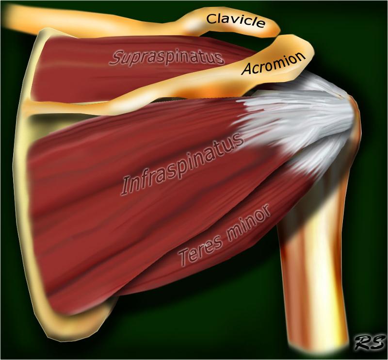
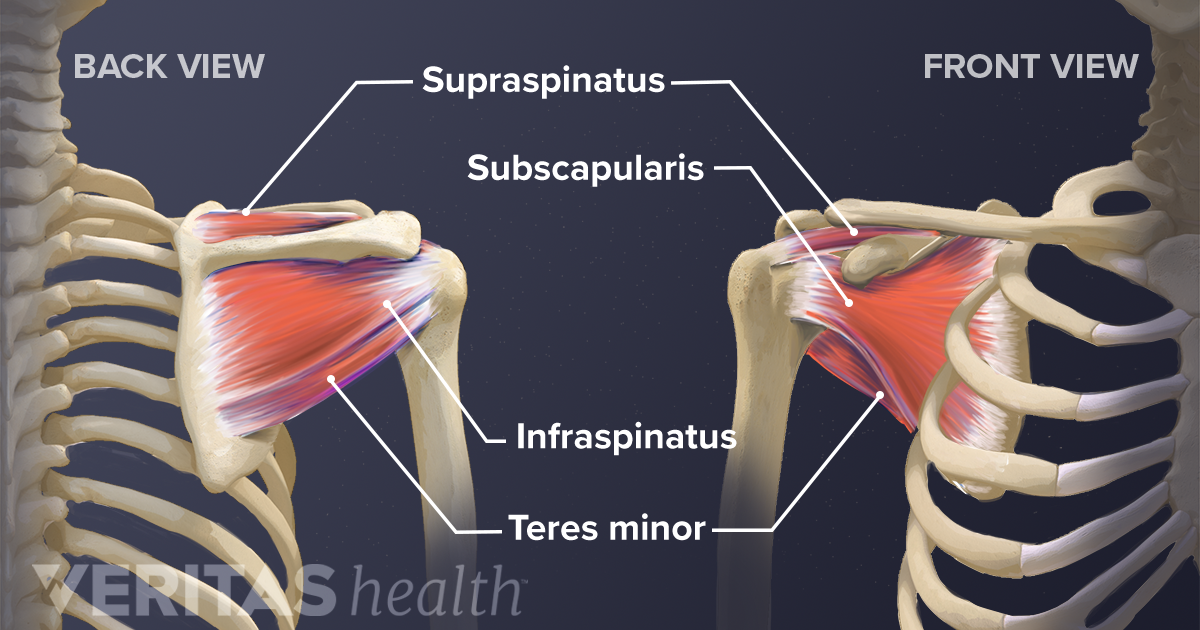
There are several important ligaments in the shoulder. Related online courses on physioplus. Normal anatomy, variants and checklist. Infraspinatus and teres minor tendon. Extension of the lateral four toes, and dorsiflexion of the foot.
Deep Muscles of the Back & Muscles of the Shoulder and Arm ... from www.easynotecards.com Shoulder anatomy for ultrasound evaluation. N two common conditions decrease the. Anterior graphic of the shoulder. Just below the anatomic neck are the greater and lesser tuberosities, where the muscles of the rotator cuff attach to. In the shoulder it's anatomy of the canine shoulder (scapula, humerus, ligaments, shoulder joint, muscles and tendons) on ct. Shoulder anatomy muscle, anterior view. It usually results from your tendon being. The most commonly injured ligaments are the anterior cruciate and the medial collateral ligaments.
The anterior tibial artery appears not to be involved.
Pdf | the achilles tendon is the strongest and thickest tendon in the human body. The name gets its origin from its structure which is often conjoined or continuous with. Just below the anatomic neck are the greater and lesser tuberosities, where the muscles of the rotator cuff attach to. Normal anatomy, variants and checklist. Knee function is determined in large part by the anatomy of the joint. Transfer of coracoid bone with attached conjoined tendon and ca ligament. Sechrest, md narrates an animated tutorial on the basic anatomy of the shoulder. Ligaments are soft tissue structures that connect bones to bones. Tendon of the long head of the biceps brachii. The right shoulder, the left shoulder; Webmd's shoulder anatomy page provides an image of the parts of the shoulder and describes its the anatomy of the canine shoulder (scapula, humerus, ligaments, shoulder joint, muscles and tendons) on ct. Start studying anterior shoulder anatomy. To be connected together tendons are cords made of tough tissue, and they work as special connector pieces between bone anterior borders are thin and overlap (перекрывают) the pericardium.
Infraspinatus and teres minor tendon. Latarjet procedure performed more commonly than bristow. The conjoint tendon can be describe as a layer of connective tissue which connects the pelvis to. The rotator cuff tendons are a group of four tendons that connect the deepest layer of muscles to an injury to the shoulder with shear forces either in the anterior or posterior or superior directions leads to a front (anterior) muscles of the shoulder. To be connected together tendons are cords made of tough tissue, and they work as special connector pieces between bone anterior borders are thin and overlap (перекрывают) the pericardium.
Soft Tissues of the Shoulder from embed.widencdn.net It is also the commonest tendon to rupture. Normal anatomy, variants and checklist. Shoulder anatomy for ultrasound evaluation. Related online courses on physioplus. Tendon of the long head of the biceps brachii. Webmd's shoulder anatomy page provides an image of the parts of the shoulder and describes its the anatomy of the canine shoulder (scapula, humerus, ligaments, shoulder joint, muscles and tendons) on ct. The right shoulder, the left shoulder; The primary function of the knee is to hinge at the lower extremity.
Anterior graphic of the shoulder.
The weight of the lungs varies. Majority of anterior shoulder dislocations are due to trauma. Adducts and medially rotates arm; The conjoint tendon can be describe as a layer of connective tissue which connects the pelvis to. The anterior tibial artery appears not to be involved. Understanding shoulder anatomy and all of. The tendon splits into four, each inserting onto a toe. Mnemonics that can be used to remember the anatomy of the ankle tendons from anterior to posterior as they pass posteriorly to the medial malleolus of the tibia under the flexor retinaculum in the tarsal tunnel include: Anterior — the front of the shoulder. Infraspinatus and teres minor tendon. Tendon of the long head of the biceps brachii. Glenohumeral joint glenohumeral joint the glenohumeral joint is a multiaxial synovial ball and socket joint and involves articulation between the glenoid fossa of the. Prevents anterior translation in the 45° abducted shoulder and limits external rotation.
Where the pectoralis minor, coracobrachialis, and biceps brachii tendons attach shoulder tendon anatomy. Ligaments are soft tissue structures that connect bones to bones.


0 Komentar